CTD-alphatech DUOTex® Coating: A Versatile Solution for Immediate Loading and Restoration
In an article titled "A Complex Case Needs Complex Pre-Planning - Part 1," published on May 11, 2010, Dr. Peter Kalitzki shares his successful experience with immediately loadable implant restoration and fixed prostheses. Faced with the challenge of placing 10 implants in the maxilla and 8 implants in the mandible, along with the patient's initial situation of a lost bite, Dr. Kalitzki emphasizes the importance of preoperative digital functional analysis and navigation scan. This case provides valuable insights into immediately loadable implant restorations, a concept that has gained significant attention from major implant manufacturers in recent years.
The CTD-alphatech DUOTex® Coating applied into the surgery, has demonstrated exceptional performance as an all-rounder solution in various dental procedures. The CTD-alphatech DUOTex® Coating applied into the surgery, has demonstrated exceptional performance as an all-rounder solution in various dental procedures. This includes immediate loading, immediate restoration, delayed immediate restoration, and conventional covered healing. The system allows for definitive bridge restorations and incorporated maxillary restorations, ensuring precise centric settings and optimized prostheses. With preoperative planning, template-guided navigation, and digital registration, the CTD-alphatech system offers enhanced possibilities for achieving long-term restorations and improved patient outcomes.
A Complex Case Needs Complex Pre-Planning
Author: Dr. Peter Kalitzki
Published on: May 11, 2010

Fig. 1 Fig. 2
Based on a patient case, the author illustrates a successful implementation of immediately loadable implant restoration with a fixed prosthesis in their practice. The procedure involved placing 10 implants in the maxilla and 8 in the mandible, considering the initial condition of the lost bite. To ensure accuracy and precision, significant emphasis was placed on preoperative digital functional analysis and navigation scan.
In this particular case, the patient required an immediately loadable implant restoration with a fixed prosthesis. Although major implant manufacturers, such as SKY Fast & Fixed (Bredent) and All-on-4 (Nobel Biocare), have actively promoted this type of restoration in recent years, it was originally developed in the 1960s and 1970s during the era of extension implants, successful cases were already reported at that time. Statistical analyses focusing on immediate care and loading have demonstrated long-term positive outcomes. Furthermore, recent animal studies on osseointegration have revealed that contact between osteocytes and the implant surface occurs much earlier than previously believed.
Prerequisites for Successful Implantation and Immediate Exposure.
Recall examinations of our patients who have had long-term implant restorations, such as bridges, primary splinted over bars, combined fixed and removable prosthetic constructions, and hybrid bridges, beyond the 10-year mark, have revealed consistent patterns:
- The length ratio between the implant body and the superstructure, ideally maintained at 1:1 or even surpassed in favour of the implant length.
- Implants with a diameter of 3.25 mm to 4 mm, designed to fit into anatomically available bone substance or adequately augmented bone.
- Maximum utilization of the implant surface to provide structural support for the superstructure.
- Presence of sufficient attached gingiva.
- Consideration of appropriate spacing between implants or between implants and the existing natural dentition to preserve the necessary biological width for the superstructure.
In the course of the recently popularized ultra-short and mini-implants, these established principles may seem outdated, but they have consistently proven their effectiveness in long term restorations. Therefore, under the aforementioned principles, Med3D navigation planning was employed for the following patient case, enabling the guided insertion of a total of 18 implants. Our practice has been utilizing the Med3D system since 2005, and it has demonstrated its reliability.
The mucosa-supported insertion template offered by the Med3D system allows for minimally invasive procedures in many cases, making it our preferred choice over the bone supported template. Additionally, the system's open nature allows for flexibility, enabling the use of different implant systems or even switching between implant systems, making it highly versatile and appealing to practitioners.
Many of our edentulous patients who have been wearing inadequate dentures for years face significant challenges. In numerous cases, their precise bite position cannot be accurately determined from a neuromuscular perspective. Consequently, emphasizing functional prosthetic reconstruction becomes even more crucial in these scenarios.
In the presented case, the existing complete dentures were initially relined to ensure optimal support. Once the prostheses were optimized, a DlR measurement was performed for functional analysis. This measurement, developed by Prof. Vogel, utilizes a digital system to determine the centricity of the mandibular condyle within the joint cavity. It focuses on the physiological extension and loading of the medial and lateral pterygoid muscles. In this specific case, the measurement showed minimal deviation. The obtained values were used to improve the occlusion of the existing prostheses and to create a wax-up for the fabrication of a moulded splint, allowing for the postoperative creation of an occlusion congruent temporary denture.
For the implant insertion, a conventional alveolar ridge incision was made. The mucosa-supported templates of the Med5D system could be accurately positioned through the remaining or reduced palatal or lingual mucosa (Fig. 1).
During the 2 mm pre-drilling, it was observed that the cancellous bone in the posterior region of the maxilla (regions 14, 15) and the anterior region of the mandible (regions 32-42) contained fat cells. In these areas, the implant sites were prepared in a subluminal manner and gradually expanded to the final implant diameter using appropriately calibrated osteotomes. This technique ensured sufficient primary stability during the surgery and promoted optimal osteocyte attachment for long-term osseointegration (Fig. 2).
In a single session, a total of 10 CTD-alphatech implants were placed in the maxilla, and 8 implants were placed in the mandible. The CTD-alphatech implant utilized in this case incorporates various design elements of a modern implant system. It features a threaded self-tapping portion in the apical region, progressive compression through decreasing thread depth in the upper third, micro rings in the cervical region, an inner cone for mechanical sealing of the implant body, a long hex rotation protection, and torsion protection in the form of three notches in the apical region.
In our opinion, an implant coating can play a role in osseointegration by acting as a barrier during insertion into existing bone structures. This coating helps remove damaged osteocytes and needs removal before the implant surface comes into contact with fresh osteocytes, facilitating the desired osseointegration. In the presented case, CTD-alphatech implants with a DUOtex® surface (roughened and etched) were used.
However, in our practice, we have achieved excellent results with DUOtex®, which is coated with a combination of hydroxyapatite, beta-tricalcium phosphate, and SiO2 using the sol-gel process. They have been successfully placed in our practice, often alongside augmentative procedures, regardless of the specific bone substitute material used.

Fig. 3: Positioned impression copings before wound closure and subsequent individual impression taking.
Diameter-congruent impression copings were initially placed on the implants (Fig. 3). Due to the presence of sufficient attached gingiva, the alveolar ridge incision could be closed almost completely, ensuring a saliva-tight environment.

Fig. 4: Positioned model implants with gingival former in the individual impression.

Fig. 5: Gingiva former positioning.
During the surgery, intraoperative impressions were taken using custom trays prepare according to the navigation tray (Fig. 4). The impression posts were replaced with 6 mm high gingiva formers (Fig. 5). The centric registration obtained from this process was used to fine-tune the occlusion and establish the bite plane.
Within 24 hours, the existing complete dentures were milled and soft-retained, allowing telescopic friction on the gingiva formers. Which not only ensures a secure fit but facilitates the shaping of the soft tissue in a manner resembling pontics.

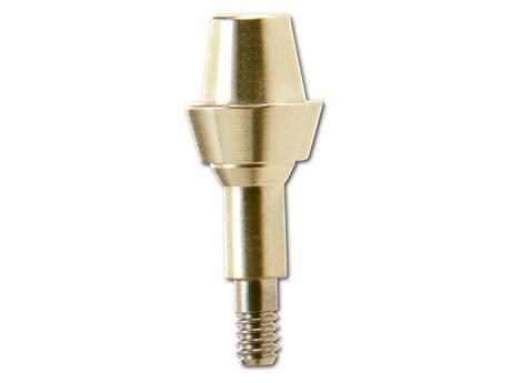
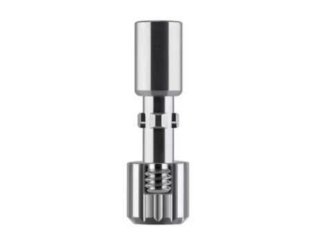
Fig. 6: Insertion key with customized abutments.
After five days, a framework try-in was performed on individualized implant abutments (Fig. 6), and the occlusion was checked. However, the soft tissue was still irritated, making it challenging to accurately assess the crown margins. A follow-up framework trial was conducted 10 days postoperatively, coinciding with the removal of sutures. At this stage, the gingiva's condition still did not allow for a precise evaluation of the red-white transitions, thus ruling out an immediate placement of the definitive restoration.
Considering the mucosal situation and the extensive bone compaction performed in 8 out of the 18 implant sites, it was decided against an immediate definitive restoration.
The existing complete dentures underwent necessary modifications to include metal reinforcement at the rim of the tooth and soft tissue relining, thus allowing the patient to have quasi-removable bridges supported by gingiva formers in both the maxilla and mandible.
The primary goal of modifying the existing prosthesis was to help the patient adapt to the new phonetic and functional conditions. Simultaneously, the implants were loaded, and open healing was utilized to shape the soft tissue in a pontic-like manner. The option of using an acetal resin long-term temporary restoration was rejected due to economic considerations in consultation with the patient.

Fig. 7: Uninflamed gingiva with a very well-defined circular shape and adequate attached gingiva.

Fig. 8: X-ray verification after the placement of the individual definitive abutments.

Fig. 9: Repeated scaffold sampling in the 11th week.
postoperative week with modeled bite points.
Subsequent X-ray checks after the placement of individualized definitive abutments showed excellent progress, with no signs of irritation and well-developed circular gingiva (Fig. 7). After another framework try-in (Fig. 8 & 9) and occlusal assessment, the splinted crownbridge dressings were definitively removed in the 12th week postoperatively then cemented into place (Figs. 10 and 11).

Fig. 10: Final bridge placement in the lower jaw after cementation.

Fig. 11: Integrated upper jaw restoration (extension bridge) immediately after cementation, with careful removal of excess cement.
In conclusion, the success of an extensive implant restoration with open healing and immediate loading relies heavily on the chosen implant system. The selected implant system must ensure adequate primary stability, even in cases where the bone density is suboptimal (as was partially the case here, with D2-D3 bone density). It is crucial to avoid excessive insertion pressure that may lead to heat necrosis.
The CTD-alphatech system with DUOtex® coating used in this case has proven to be a versatile solution suitable for immediate loading, immediate restoration, delayed immediate restoration, and conventional covered healing in our practice.
When restoring the dentition of edentulous patients or those with total prostheses, the absence of neuromuscular feedback through tactile receptors in the periodontiumm necessitates the implementation of functional analysis measures, these measures should be carried out not only during the definitive prosthetic restoration but also before the surgical procedure. The utilization of the DIR system for registration ensures a physiologically centred occlusion, allowing the optimization of existing prostheses, as seen in the present case, or the fabrication of new interim prostheses in the case of
different initial conditions. By acclimating the patient to the new physiologically centred occlusion, the transfer of the centric registration obtained under physiological muscle loading ensures that the implant-supported fixed denture is not only more readily accepted by the patient but avoids overloading caused by misalignment.
Preoperative planning for the application of template-guided navigation, along with the incorporation of digital registration, offers the opportunity to further optimize the superstructures to be integrated. This facilitates the achievement of long-term restorations for our patients with greater ease.


